
Heart Failure with Preserved EF (HFpEF)
Stiff Ventricle · Raised Filling Pressures · The Comorbidity-Driven Epidemic
Heart failure with preserved ejection fraction — HFpEF — is defined by heart failure symptoms alongside a left ventricular ejection fraction of 50% or higher. The pump squeezes normally. The problem is that it does not relax normally, and the stiff ventricle demands abnormally high filling pressures to accept blood. Those pressures back up into the lungs, and the patient becomes breathless.[1] HFpEF now accounts for roughly half of all heart failure cases in developed countries, and that fraction is rising. It strikes older women with hypertension, obesity, diabetes, and atrial fibrillation more than any other group. For decades it was the most treatment-resistant major cardiovascular syndrome in medicine — trial after trial of renin-angiotensin blockade returned negative. That changed in 2021, when SGLT2 inhibitors became the first drug class to reduce hospitalizations in this population. Understanding HFpEF means understanding why a normal ejection fraction can coexist with severe disability — and why comorbidities, not a failing pump, are the engine of the disease.[1]
For most of the 20th century, clinicians spoke of heart failure as if it were a single entity. A failing heart was one that could no longer pump adequately — systolic dysfunction, dilated cardiomyopathy, the reduced ejection fraction that Starling's experiments in the 1890s had taught everyone to fear. The idea that a ventricle with preserved contractile function could produce the same clinical syndrome of congestion, breathlessness, and exercise intolerance barely registered as a distinct problem. The phrase "diastolic heart failure" began appearing in the literature in the 1980s, largely through investigators trying to explain why so many hospitalized patients with textbook heart failure had normal echocardiographic systolic function. It was not until the late 1990s that the term "heart failure with preserved ejection fraction" took hold — and with it, the recognition that this was a distinct pathophysiologic entity, not merely mild systolic dysfunction missed on a crude study.[1]
The therapeutic history of HFpEF is, bluntly, a history of failure. Every drug class that worked brilliantly in heart failure with reduced ejection fraction was tried in HFpEF and failed. CHARM-Preserved (2003) tested candesartan, an angiotensin receptor blocker, in 3,023 patients with ejection fraction above 40%.[2] The primary endpoint — cardiovascular death or heart failure hospitalization — was not significantly reduced. I-PRESERVE (2008) enrolled 4,128 patients with ejection fraction of 45% or higher to test irbesartan.[3] Another negative result. By the early 2010s, therapeutic nihilism had settled over the field. Some began to wonder whether HFpEF was simply too heterogeneous — too many different diseases wearing the same clinical mask — to respond to any single intervention.
Then came TOPCAT. The Treatment of Preserved Cardiac Function Heart Failure with an Aldosterone Antagonist trial randomized 3,445 patients to spironolactone or placebo and reported its primary results in 2014.[4] The composite of cardiovascular death, aborted cardiac arrest, and heart failure hospitalization was not significantly reduced. But what came next changed the interpretation of that trial entirely. Post-hoc metabolite analysis revealed that approximately 30% of patients enrolled in Russia and Georgia had undetectable blood levels of canrenone — the active breakdown product of spironolactone — suggesting they had never taken the drug. Among patients enrolled in the Americas, spironolactone reduced the primary composite with a hazard ratio of 0.82. In Eastern Europe, the hazard ratio was 1.10. The drug had not failed. The trial had been contaminated by nonadherence. PARAGON-HF (2019) then tested sacubitril-valsartan — the ARNI that transformed HFrEF — in nearly 4,800 HFpEF patients.[5] It narrowly missed its primary endpoint (p=0.06), though subgroup analyses suggested possible benefit in women and patients with ejection fractions in the 45–57% range.
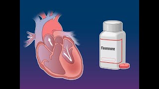
The era of therapeutic nihilism ended with EMPEROR-Preserved (2021), which showed that empagliflozin — a sodium-glucose cotransporter 2 inhibitor first developed for diabetes — reduced the composite of cardiovascular death and heart failure hospitalization by 21% in patients with HFpEF.[6] DELIVER (2022) confirmed the class effect with dapagliflozin, reducing the same composite by 18%.[7] FINEARTS-HF (2024) then showed that finerenone, a non-steroidal mineralocorticoid receptor antagonist, reduced total worsening heart failure events and cardiovascular death by 16% compared with placebo.[8] After four decades, HFpEF finally had treatments backed by class I evidence.
The fundamental defect in HFpEF is impaired myocardial relaxation — what cardiologists call diastolic dysfunction. Relaxation is an active, energy-requiring process. After each contraction, the myocardium must actively pump calcium out of the cytoplasm so the sarcomere can lengthen and the ventricle can accept blood. When energy supply is impaired — as it is in hypertension, obesity, or ischemia — calcium removal slows, the chamber takes longer to lengthen, and the ventricle becomes stiff. This stiffness is described by a steep pressure-volume relationship: a small increase in filling volume generates a disproportionately large increase in filling pressure. The patient's lungs detect this pressure surge and respond with dyspnea.[1]
Comorbidities are not bystanders in HFpEF. They are the drivers. Hypertension promotes concentric left ventricular hypertrophy — increased wall thickness that further stiffens the chamber. Obesity generates systemic inflammation and neurohormonal activation, increases intrathoracic pressure, and expands circulating blood volume, all of which raise filling pressures. Diabetes causes myocardial fibrosis through advanced glycation end products and intracellular lipid accumulation. Atrial fibrillation, which is both cause and consequence of elevated left atrial pressure, eliminates the atrial "kick" that normally contributes 20–30% of diastolic filling — a loss the stiff ventricle cannot compensate for. The heart in HFpEF has not failed in the conventional sense. It has been remodeled into inflexibility by the metabolic environment surrounding it.[1]
The hemodynamic signature of HFpEF is most apparent during exercise. At rest, many confirmed HFpEF patients have only mildly elevated filling pressures. During exertion, cardiac output must rise, but the stiff ventricle can only increase output by raising filling pressure further — the only mechanism available when heart rate rises and diastolic filling time shortens. Pulmonary capillary wedge pressure may be normal at rest (less than or equal to 12 mmHg) but spike above 25 mmHg with modest exercise. This is why exercise right heart catheterization remains the gold standard for diagnosing HFpEF when resting hemodynamics are ambiguous. Clinically, it explains the cardinal symptom: exertional dyspnea out of proportion to any resting finding.
At the cellular level, HFpEF is characterized by increased collagen cross-linking, reduced expression of titin (the giant elastic protein that acts as a molecular spring in the sarcomere) in its more compliant N2BA isoform, and impaired nitric oxide signaling. Obesity-driven inflammation elevates circulating cytokines that reduce myocardial nitric oxide bioavailability, impairing both relaxation and the vasodilatory reserve needed for exercise. This microvascular inflammatory model reframes HFpEF as a systemic metabolic syndrome with cardiac end-organ damage — which is precisely why drugs targeting systemic pathways (SGLT2 inhibitors, GLP-1 agonists, mineralocorticoid antagonists) are showing the most promise.
HFpEF is not one disease. Recognizing the predominant phenotype in a given patient shapes management. The most commonly described subtypes share the same hemodynamic signature but arrive there through different mechanisms.[1]
Obese HFpEF
BMI typically above 35 kg/m². Massive pericardial fat compresses the ventricle from outside. Elevated intrathoracic pressure from abdominal obesity raises cardiac filling pressures even at rest. BNP is disproportionately low for disease severity. Weight loss — including with tirzepatide (SUMMIT trial) — dramatically improves symptoms and functional capacity.
Hypertensive HFpEF
Concentric LV hypertrophy, small LV cavity, preserved systolic function. Long-standing pressure overload from uncontrolled hypertension. Usually the older woman with decades of borderline BP readings. Excellent BP control is the cornerstone of therapy — the LV will partially reverse hypertrophy with sustained load reduction over months to years.
Atrial Fibrillation HFpEF
AF and HFpEF create a vicious cycle: elevated left atrial pressure begets AF, and AF eliminates the atrial contribution to diastolic filling that the stiff LV depends upon. Rhythm control is increasingly preferred over rate control in symptomatic patients. Loss of the atrial kick can precipitate acute decompensation even at modestly rapid ventricular rates.
CKD / Cardiorenal HFpEF
Chronic kidney disease amplifies volume retention, neurohormonal activation, and anemia. Diuretic resistance is common. SGLT2 inhibitors simultaneously improve cardiac and renal outcomes in this subgroup — one of their most clinically important attributes. Loop diuretics may need supplementation with thiazide-type agents to overcome nephron adaptation.
The most common triggers of acute HFpEF decompensation are atrial fibrillation with rapid ventricular response, dietary sodium excess, missed diuretic doses, uncontrolled hypertension, and infection. Unlike HFrEF, the HFpEF patient decompensates from a sudden rise in filling pressure, not a fall in cardiac output. Blood pressure is often elevated or normal at presentation — not low. Hypertensive emergency presenting as acute pulmonary edema in an older woman with preserved EF is classic HFpEF decompensation — treat the hypertension aggressively with IV nitrates and loop diuretics. Avoid aggressive vasodilation in the rare hypotensive HFpEF patient. The small, stiff LV is preload-dependent; overdiuresis can precipitate hypotension and prerenal azotemia just as readily as underdiuresis causes congestion.
HFpEF is a clinical diagnosis of exclusion as much as inclusion. The exam findings reflect elevated filling pressures, not reduced cardiac output — the hands are warm, the pulse volume is normal, but the jugular venous pressure is elevated and the lungs may be wet. The absence of an S3 gallop does not exclude the diagnosis. A prominent S4 — the sound of atrial contraction into a stiff ventricle — is common in HFpEF and often absent in HFrEF, where the atrium is too dilated to generate a crisp sound.[1]
| Finding | Sensitivity | Specificity | Clinical Pearl |
|---|---|---|---|
| Elevated JVP | ~70% | ~80% | Best exam sign of elevated RA pressure; hardest to assess in obesity — POCUS of the internal jugular is more reliable |
| S4 gallop | ~60% | ~80% | Atrial contraction into stiff LV; heard best at apex with the bell of the stethoscope |
| Bilateral leg edema | ~50% | Low | Non-specific; venous insufficiency and medication effects confound in older patients |
| Bibasilar crackles | ~50% | Moderate | Often absent in chronic HFpEF due to lymphatic adaptation; reliably present in acute decompensation |
| Absent displaced PMI | Low | High | Non-dilated LV in HFpEF keeps the PMI at the apex; a laterally displaced PMI should raise suspicion for HFrEF instead |
| H2FPEF score ≥6 | ~80% | ~85% | Points: Heavy (obesity) +2, Hypertensive (≥2 meds) +1, AF +3, Pulmonary HTN +1, Elder (age >60) +1, Filling pressure (E/e' >9) +1. Score 0–1 = low probability; ≥6 = high |
The H2FPEF score — standing for Heavy, Hypertensive, AF, Pulmonary hypertension, Elder, and elevated Filling pressure — was derived and validated in a cohort of 414 patients and provides a practical clinical tool to distinguish HFpEF from non-cardiac dyspnea without requiring invasive hemodynamics.[9] BNP interpretation requires caution: obesity markedly suppresses natriuretic peptide levels. A BNP of 100 pg/mL in a 350-pound patient may represent the same hemodynamic burden as a BNP of 400 pg/mL in a normal-weight patient. The H2FPEF score implicitly accounts for obesity by treating it as a positive weighted variable rather than a reason to discount a low BNP.
In HFpEF, echocardiography is both the primary diagnostic tool and the monitoring instrument. The focused bedside exam addresses: (1) Is the LV non-dilated with preserved systolic function? (2) Is the left atrium enlarged — a durable marker of chronically elevated filling pressure? (3) Is there evidence of elevated filling pressure right now, via E/e' and B-line count? (4) Is the interventricular septum thickened, suggesting hypertensive or infiltrative remodeling?
E/e' ratio: Place the pulsed-wave Doppler sample volume at the mitral leaflet tips in the apical 4-chamber view to measure the E wave — the early mitral inflow velocity representing the pressure gradient from left atrium to left ventricle at the start of diastole. Then use tissue Doppler imaging at the septal and lateral mitral annulus to measure e' — the early diastolic annular velocity, which reflects myocardial relaxation rate. The ratio E/e' correlates with mean left atrial pressure. Average E/e' above 15 strongly suggests elevated LV filling pressure (specificity approximately 85%). Values below 8 effectively exclude it. The 8–14 range is indeterminate and requires additional echo parameters.
B-lines on lung ultrasound: Scan the anterior and lateral chest wall using a phased-array or curvilinear probe. B-lines — vertical hyperechoic artifacts arising from the pleural line and extending to the screen's bottom without fading — represent interstitial edema from elevated pulmonary capillary pressure. Three or more B-lines per zone in multiple bilateral zones indicates pulmonary congestion with high sensitivity for elevated filling pressures. B-line counts correlate with intracardiac pressures and fall dynamically with effective diuresis — making lung ultrasound an excellent monitoring tool throughout hospitalization.[10]
Left atrial size: In the parasternal long-axis view, measure LA anteroposterior diameter. An LA diameter above 4.0 cm in women or 4.5 cm in men (or LA volume index above 34 mL/m²) reflects chronic exposure to elevated filling pressures. LA enlargement is one of the four standard criteria for diastolic dysfunction grading and — combined with E/e' and tricuspid regurgitation velocity — defines the 2016 ASE/EACVI diastolic dysfunction algorithm. Unlike E/e', LAVI reflects the cumulative hemodynamic burden over months, not just the current moment.
The laboratory workup in HFpEF establishes the diagnosis, quantifies disease severity, identifies comorbidities, and guides therapy. No single test confirms the diagnosis alone; the picture comes from integrating multiple parameters and the clinical story.[1]
| Test | What It Tells You | Key Interpretation Points |
|---|---|---|
| BNP / NT-proBNP | Marker of myocardial wall stress and filling pressure | Obesity suppresses BNP substantially — a BNP of 100 pg/mL in a 350-lb patient may represent equivalent hemodynamic burden to a BNP of 400 in a normal-weight patient. NT-proBNP above 125 pg/mL supports HF diagnosis. Use clinical context and H2FPEF score alongside the biomarker. |
| Echocardiographic E/A ratio | Pattern of mitral inflow reflecting diastolic filling dynamics | Grade I: E/A <0.8 (impaired relaxation — slow, sluggish early filling). Grade II: E/A 0.8–2.0 with E/e' >14 or LA enlargement (pseudonormal — appears normal but filling pressures are elevated). Grade III: E/A >2 with short deceleration time (restrictive — very high filling pressures, advanced disease). |
| E/e' ratio | Noninvasive surrogate for LV filling pressure | Average E/e' above 15 = elevated filling pressure (specificity ~85%). Below 8 = normal (sensitivity ~85%). The 8–14 zone is indeterminate — combine with LA volume and TR velocity to grade diastolic function. |
| LA volume index (LAVI) | Durable marker of chronically elevated left atrial pressure | LAVI above 34 mL/m² is abnormal and reflects the time-integrated burden of elevated filling pressures over months. Essential for diastolic dysfunction grading. Unlike acute E/e', LAVI does not normalize overnight with diuresis. |
| Deceleration time (DT) | Speed of early mitral inflow deceleration after peak E wave | DT <150 ms suggests restrictive physiology and high filling pressures. DT >220 ms with low E/A ratio suggests Grade I (impaired relaxation). Normal 150–220 ms. |
| Cardiac MRI | Tissue characterization: fibrosis, infiltration, pericardial disease | Late gadolinium enhancement detects focal fibrosis. T1 mapping and extracellular volume fraction (ECV) quantify diffuse interstitial fibrosis. MRI is the gold standard when cardiac amyloid, sarcoid, hemochromatosis, or constrictive pericarditis is in the differential — all of which can mimic HFpEF. |
| Exercise right heart catheterization | Gold standard for elevated filling pressures with exertion | PCWP ≥25 mmHg with exercise in a patient with exertional dyspnea and preserved EF confirms HFpEF when noninvasive data are equivocal. Resting wedge pressure may be normal in established HFpEF — exercise hemodynamics unmask the true physiology. |
Treatment of HFpEF follows two parallel strategies: symptom relief through decongestion, and modification of the comorbidity burden that drives disease progression. SGLT2 inhibitors are now the only drug class with class IIa evidence and Level B-R data for reducing hospitalizations in HFpEF, supported by the 2022 ACC/AHA/HFSA guideline.[11]
| Intervention | When & How | Evidence & Key Points |
|---|---|---|
| SGLT2 inhibitors (empagliflozin 10 mg or dapagliflozin 10 mg daily) | First-line in all HFpEF patients without contraindication. Start regardless of diabetes status. Maintain as long as tolerated (eGFR threshold: ≥20 mL/min/1.73m² for both). | EMPEROR-Preserved: 21% RRR in CV death + HF hospitalization (HR 0.79, p<0.001)[6]; DELIVER: 18% RRR (HR 0.82, p<0.001)[7]. Benefits consistent across EF subgroups, diabetes status, sex, and background therapy. Class IIa, Level B-R per 2022 guidelines.[11] |
| Loop diuretics (furosemide, torsemide, bumetanide) | For congestion relief in volume-overloaded patients. Titrate to symptom relief and stable dry weight. Avoid excessive diuresis — the stiff LV is preload-sensitive. | No mortality data; symptomatic benefit is the primary goal. Torsemide has more predictable bioavailability than furosemide. Monitor electrolytes and renal function. Overdiuresis in a small, hypertrophied LV can cause paradoxical hypotension and prerenal azotemia. Class I recommendation.[11] |
| Finerenone (non-steroidal MRA, 10–40 mg daily) | Consider in patients with EF ≥40%, elevated NT-proBNP, and eGFR ≥25 mL/min. Titrate to 20–40 mg if potassium remains below 5.0 mEq/L. | FINEARTS-HF (2024): 16% RRR in total worsening HF events + CV death (rate ratio 0.84, p=0.007)[8]. First MRA with a positive primary endpoint in HFpEF. Lower hyperkalemia risk than steroidal MRAs at equivalent efficacy. Not yet in 2022 guidelines — emerging evidence. |
| Blood pressure control (target <130/80 mmHg) | Treat to guideline targets. ACE inhibitors, ARBs, and thiazide diuretics are all acceptable. Beta-blockers for rate control in AF. Avoid dihydropyridine CCBs in patients with AF who need rate control (less effective). | Hypertension is the most prevalent and modifiable risk factor for HFpEF. Reversing LV concentric hypertrophy requires years of sustained BP control. CHARM-Preserved[2] and I-PRESERVE[3] were negative for primary endpoints but support BP lowering as physiologically rational. Class I recommendation.[11] |
| AF management (rhythm control preferred) | Rhythm control preferred over rate control in younger, symptomatic patients. Early cardioversion if HFpEF decompensation is precipitated by AF. Rate target ≤80 bpm at rest if rate control is chosen. | The stiff ventricle of HFpEF relies on the atrial contribution to filling. Loss of atrial kick in AF can precipitate acute decompensation. Early rhythm control has demonstrated cardiovascular benefit in patients with AF and HF. Class I recommendation for management of AF in HFpEF.[11] |
| Obesity treatment (structured weight loss, GLP-1 / dual agonists) | Target sustained weight loss. Tirzepatide (GLP-1/GIP dual agonist) and semaglutide (GLP-1 agonist) have shown dramatic symptom improvement in obese HFpEF patients. Bariatric surgery may be considered in severe obesity. | SUMMIT trial (tirzepatide): 15-point improvement in KCCQ score and significant reduction in hierarchical composite endpoint in obese HFpEF patients. Weight loss reduces pericardial fat, intrathoracic pressure, and systemic inflammation — directly addressing the dominant pathophysiologic drivers in the obese phenotype. |
| Exercise training | Structured aerobic training, 3–5 sessions per week, 30–45 minutes. Cardiac rehabilitation if available. Begin after clinical stability is achieved. | Exercise training improves peak VO2, six-minute walk distance, and quality of life through peripheral oxygen extraction and skeletal muscle conditioning. No mortality data available. Recommended as adjunct therapy in all clinically stable patients. Class IIa recommendation.[11] |
| Sacubitril-valsartan (ARNI, 24/26–97/103 mg twice daily) | Consider in HFpEF with EF 45–55%, particularly women and those with lower-preserved EF. Not appropriate for patients already on ACE inhibitor (36-hour washout required). | PARAGON-HF: narrowly missed primary endpoint (RR 0.87, 95% CI 0.75–1.01, p=0.06)[5]. Subgroup analyses suggest benefit in women and patients with EF in the low-preserved range. ACC/AHA Class IIb, Level B-R.[11] |
| Recommendation — ACC/AHA/HFSA 2022 Guideline[11] | Class | Level of Evidence |
|---|---|---|
| SGLT2 inhibitors to reduce HF hospitalizations and CV mortality in patients with HFpEF | IIa | Level B-R |
| Diuretics to relieve congestion and improve symptoms in HFpEF | I | Level C |
| Management of hypertension and atrial fibrillation in HFpEF to reduce symptoms and prevent hospitalizations | I | Level C |
| MRA (spironolactone) may be considered to decrease hospitalizations in HFpEF | IIb | Level B-R |
| ARBs may be considered to decrease hospitalizations in HFpEF | IIb | Level B-R |
| ARNI (sacubitril-valsartan) may be considered in select HFpEF patients, particularly women and those with lower-preserved EF | IIb | Level B-R |
| Routine use of nitrates or phosphodiesterase-5 inhibitors to improve activity tolerance in HFpEF is not recommended | III: No Benefit | Level B-R |
TOPCAT enrolled patients across two geographic regions: the Americas (United States, Canada, Brazil, Argentina) and Eastern Europe (Russia and Georgia). When the results were published in 2014,[4] the overall trial was negative — spironolactone did not significantly reduce the composite primary endpoint of cardiovascular death, aborted cardiac arrest, or heart failure hospitalization. But the geographic heterogeneity was striking and deeply suspicious: the hazard ratio in the Americas was 0.82 (95% CI 0.69–0.98), clearly favoring spironolactone. In Russia and Georgia, it was 1.10 (95% CI 0.79–1.51) — going in the opposite direction.
Investigators went looking for an explanation. They measured canrenone — the active circulating metabolite that spironolactone is converted to in the liver, which serves as pharmacokinetic proof that the drug was ingested. Among Russian patients randomized to spironolactone, approximately 30% had undetectable canrenone levels. In the Americas, only 3% were undetectable. Meanwhile, hyperkalemia — an expected and reliable pharmacodynamic consequence of mineralocorticoid receptor blockade — occurred 3.5-fold more often in the spironolactone arm than placebo in the Americas, exactly as expected. In Russia and Georgia, hyperkalemia rates were similar in both arms. The drug appeared never to have been taken by a large fraction of Eastern European patients.
Further irregularities emerged. Event rates in the Eastern European placebo group were implausibly low by any comparison to epidemiological data. The baseline characteristics of Russian enrollees did not match the expected HFpEF phenotype. Multiple investigations raised concerns about enrollment practices, data integrity, and site conduct. The NHLBI acknowledged the regional discrepancy but raw patient-level data from Russia were not available for full adjudication.
The clinical implication is significant: if TOPCAT is restricted to the Americas-only population — where pharmacokinetic evidence confirms the drug was actually taken — spironolactone reduced heart failure hospitalizations significantly. Many cardiologists have concluded that spironolactone works in HFpEF, that TOPCAT was corrupted by large-scale nonadherence in Eastern Europe, and that the apparent negative overall result reflects a trial integrity failure, not a drug failure. The ACC/AHA 2022 guideline assigned a Class IIb recommendation to MRAs in HFpEF[11] — a cautious acknowledgment that the drug may work, muddied by a corrupted trial. FINEARTS-HF (2024) has now provided a clean positive signal for the MRA class with finerenone,[8] which many interpret as the definitive vindication of mineralocorticoid blockade in HFpEF — even if TOPCAT never delivered a clean answer about spironolactone specifically.
Empagliflozin reduced CV death and HF hospitalization by 21% in patients with EF above 40%. The first positive outcome trial in HFpEF.
Dapagliflozin reduced the primary composite by 18% in HFpEF and HFmrEF (EF above 40%). Confirmed the SGLT2 inhibitor class effect across the full EF spectrum.
Finerenone reduced total worsening HF events and CV death by 16% in HFpEF and HFmrEF. First positive primary endpoint for the MRA class in this population.
Derivation and validation of the H2FPEF clinical score for diagnosing HFpEF without invasive hemodynamics. AUC 0.841 in the validation cohort.

[1] Borlaug BA, Redfield MM. Chapter 49: Diagnosis and Management of Heart Failure with Preserved Ejection Fraction. In: Fuster V, et al., eds. Hurst's The Heart, 15th ed. McGraw-Hill, 2023 (Updated April 2023).
[2] Yusuf S, et al. Effects of candesartan in patients with chronic heart failure and preserved left-ventricular ejection fraction: the CHARM-Preserved Trial. Lancet. 2003;362(9386):777–781. PMID: 13678871. [PubMed]
[3] Massie BM, et al. Irbesartan in patients with heart failure and preserved ejection fraction (I-PRESERVE). N Engl J Med. 2008;359(23):2456–2467. PMID: 19001508. [PubMed]
[4] Pitt B, et al. Spironolactone for heart failure with preserved ejection fraction (TOPCAT). N Engl J Med. 2014;370(15):1383–1392. PMID: 24716680. [PubMed]
[5] Solomon SD, et al. Angiotensin-neprilysin inhibition in heart failure with preserved ejection fraction (PARAGON-HF). N Engl J Med. 2019;381(17):1609–1620. PMID: 31475794. [PubMed]
[6] Anker SD, et al. Empagliflozin in heart failure with a preserved ejection fraction (EMPEROR-Preserved). N Engl J Med. 2021;385(16):1451–1461. PMID: 34449189. [PubMed]
[7] Solomon SD, et al. Dapagliflozin in heart failure with mildly reduced or preserved ejection fraction (DELIVER). N Engl J Med. 2022;387(12):1089–1098. PMID: 36027570. [PubMed]
[8] Solomon SD, et al. Finerenone in heart failure with mildly reduced or preserved ejection fraction (FINEARTS-HF). N Engl J Med. 2024;391(16):1475–1485. PMID: 39225278. [PubMed]
[9] Reddy YNV, et al. A simple, evidence-based approach to help guide diagnosis of heart failure with preserved ejection fraction (H2FPEF Score). Circulation. 2018;138(9):861–870. PMID: 29792299. [PubMed]
[10] Platz E, et al. Lung ultrasound in acute heart failure: prevalence of pulmonary congestion and short- and long-term outcomes. JACC Heart Fail. 2019;7(10):849–858. PMID: 31582107. [PubMed]
[11] Heidenreich PA, et al. 2022 AHA/ACC/HFSA Guideline for the Management of Heart Failure. J Am Coll Cardiol. 2022;79(17):e263–e421. PMID: 35363499. [PubMed]