Pulmonology · Interstitial & Diffuse Parenchymal Lung Disease
Cryptogenic Organizing Pneumonia (COP)
Intra-Alveolar Fibroblast Plugs · Steroid-Responsive · High Relapse Risk
Cryptogenic organizing pneumonia (COP) is an acute-to-subacute idiopathic interstitial pneumonia defined by intra-alveolar plugs of granulation tissue — so-called Masson bodies — filling the distal airways, alveolar ducts, and alveoli without a definable inciting cause.[1] It is the most steroid-responsive of the idiopathic interstitial pneumonias, with dramatic clinical and radiographic improvement in the majority of patients within weeks of starting prednisone — an outcome that stands in sharp contrast to idiopathic pulmonary fibrosis, where corticosteroids are actively harmful. The practical problem with COP is not that it is hard to treat; it is that it is almost always diagnosed late. Most patients receive two, three, or four courses of antibiotics for presumed community-acquired pneumonia before anyone thinks to look beyond infection as the cause of subpleural consolidations that refuse to resolve.[2] Once recognized, however, the majority of patients respond rapidly to corticosteroids — though relapse rates of 13–58% on steroid tapering mean that treatment is rarely a single short course.
The condition we now call COP was first described in 1983 by Davison, Heard, McAllister, and Turner-Warwick, who reported eight patients with histological intra-alveolar organization and no identifiable infectious or environmental trigger.[3] These patients shared a striking clinical pattern: a short history of severe dyspnea, cough, malaise, weight loss, bilateral radiographic shadowing, and a markedly elevated erythrocyte sedimentation rate. All were dramatically responsive to prednisolone, but all relapsed rapidly as the dose was reduced — a clinical signature that has defined the disease ever since. Davison's group proposed the term "cryptogenic organizing pneumonitis" to distinguish the idiopathic form from the post-infectious organizing pneumonia that pathologists had been describing for decades. The word "cryptogenic" — meaning of unknown origin — was a deliberate choice to signal that the process had no identifiable driver, and that the clinician's obligation was first to exclude every possible cause before accepting the diagnosis as idiopathic.
Two years later, in 1985, Gary Epler and colleagues at the Massachusetts General Hospital published a landmark series of 50 patients in the New England Journal of Medicine under the term "bronchiolitis obliterans organizing pneumonia," or BOOP.[4] The Epler paper was larger, more systematically characterized, and appeared in the highest-impact journal in medicine — and as a result, BOOP became the dominant term in North American pulmonology for the next two decades. The Epler paper established what has remained the clinical fingerprint of the disease: a subacute flu-like illness in a middle-aged patient with bilateral consolidations on chest imaging, no pathogen on culture or bronchoscopy, and dramatic steroid responsiveness. Of the 50 patients in Epler's original series, 94% improved or recovered on corticosteroids, establishing the paradigm for treatment that remains largely unchanged today.
The terminology problem was resolved — or at least formalized — in 2002, when the American Thoracic Society and European Respiratory Society published the first multinational classification of the idiopathic interstitial pneumonias. The 2002 statement officially replaced "BOOP" with "cryptogenic organizing pneumonia" (COP) as the preferred term, reserving "bronchiolitis obliterans syndrome" (BOS) for the obstructive airway disease seen in lung transplant rejection — a condition that shares no meaningful pathophysiology with COP despite the name overlap.[5] COP was classified as an acute/subacute IIP, distinct from the chronic fibrosing IIPs (IPF, NSIP) and the smoking-related IIPs (DIP, RB-ILD). The 2013 ATS/ERS update to this classification reinforced COP's position in the IIP framework, noting its distinctive clinical, radiographic, and histopathologic features and its generally favorable prognosis when treated.[5]
The broader category of organizing pneumonia — the pathologic pattern, not just the idiopathic form — was recognized throughout the 20th century in the setting of infections, drug reactions, connective tissue diseases, and radiation injury. What Davison and Epler contributed was the understanding that this pathologic process could occur without any identifiable trigger, and that in this idiopathic form, corticosteroids could reverse what appeared radiographically to be fixed consolidation. This insight set COP apart not only clinically but conceptually: unlike the fibrotic IIPs where the lung heals with scar, in COP the intra-alveolar plugs of granulation tissue can dissolve, the alveolar architecture can be preserved, and the patient can return to near-normal lung function — provided the diagnosis is made before chronic remodeling sets in.
COP is an intra-alveolar rather than interstitial disease, a distinction that matters both pathologically and therapeutically. The defining lesion is the Masson body — a polypoid plug of granulation tissue composed of myofibroblasts embedded in a loose connective tissue matrix, filling the lumen of a respiratory bronchiole, alveolar duct, or alveolus. These plugs form as a stereotyped response to injury of the alveolar epithelium: the injured type I pneumocyte is replaced by proliferating type II cells; inflammatory cells (macrophages, lymphocytes, plasma cells) accumulate; and fibroblasts migrate into the alveolar space and proliferate, depositing provisional connective tissue. The key distinguishing feature of COP is that this fibroblastic response remains loose and myxoid rather than progressing to dense collagen deposition. This loose connective tissue matrix — not yet organized into rigid collagen fibers — is susceptible to dissolution by corticosteroids, which suppress fibroblast proliferation, reduce inflammatory cytokines (particularly TGF-β, IL-6, and TNF-α), and allow the alveolar architecture to re-expand.[1]
In contrast to IPF — where the primary lesion is aberrant epithelial wound healing driving progressive fibrosis independent of ongoing inflammation — COP is driven by a sustained inflammatory stimulus that the immune system cannot resolve on its own. Whether that stimulus is a viral infection, an autoimmune process, a drug reaction, or truly no identifiable cause (idiopathic COP), the pathologic response in the distal airspace is the same: organizing granulation tissue filling the alveolar lumen while the septal architecture remains relatively intact. This preservation of architecture explains why COP can be completely reversible with treatment — there is no honeycombing, no fibroblastic foci, no irreversible structural destruction of the kind seen in UIP/IPF. BAL in COP shows a characteristic pattern of mixed cellularity — lymphocytes, neutrophils, and eosinophils are all elevated, reflecting the diverse inflammatory response — rather than the neutrophilic predominance of bacterial pneumonia or the pure lymphocytosis of hypersensitivity pneumonitis.[2]
The propensity for relapse — seen in 13–58% of patients during steroid tapering — reflects the underlying biology: in many patients, the inciting stimulus is not fully eliminated by steroid-mediated suppression, and partial resolution during treatment is followed by recurrence when the anti-inflammatory brake is removed. In secondary COP (associated with CTD, drugs, or infection), the relapse risk is particularly high if the underlying cause has not been treated. In idiopathic COP, relapse typically responds to re-escalation of corticosteroids and does not carry the same grim prognosis as an acute exacerbation of IPF. Relapses in COP are annoying and require prolonged treatment, but they are not fatal — a crucial distinction.
| Category | Common Triggers / Associations | Key Features | Prognosis / Course |
|---|---|---|---|
| Idiopathic COP | No identifiable cause; diagnosis of exclusion | Classic presentation: subacute flu-like illness, bilateral subpleural consolidations, reverse halo sign on CT. Responsive to prednisone. No associated CTD, drug, or infection.[6] | Generally excellent — >80% 5-year survival. Relapse rate 13–58%. Relapses respond to re-treatment. Death from COP alone is rare. |
| CTD-associated OP | Polymyositis / dermatomyositis (most common), RA, systemic sclerosis, Sjögren syndrome, SLE, MCTD | OP may be the initial presentation of CTD. Serology (ANA, RF, anti-CCP, myositis-specific antibodies: anti-Jo-1, anti-MDA5) essential. Co-existing NSIP or UIP pattern may be present.[7] | Longer treatment required; relapse common if underlying CTD is not controlled. Prognosis tied to severity of underlying CTD. |
| Drug-induced OP | Amiodarone, nitrofurantoin, bleomycin, methotrexate, phenytoin, sulfasalazine, checkpoint inhibitors (anti-PD-1/PD-L1) | Drug history is critical — OP pattern on biopsy in the setting of a known offending agent. Requires discontinuation of causative drug. May co-exist with direct drug toxicity (NSIP pattern). | Good if drug is stopped promptly. Checkpoint inhibitor-associated OP typically steroid-responsive but may require high-dose therapy. |
| Post-infectious OP | Influenza A (H1N1), COVID-19, Mycoplasma, Legionella, Pneumocystis, bacterial pneumonia (non-resolving) | Organizing pneumonia pattern persists after the infectious agent has been cleared. Distinguishable from active infection by negative cultures/PCR and failure to respond to additional antibiotics. | Generally self-limited or steroid-responsive. COVID-19-associated OP may require prolonged treatment. |
| Post-radiation OP | Breast cancer radiation (3–6 months after treatment), thoracic radiation for any malignancy | Bilateral, migratory consolidations beyond the radiation field — distinguishes from classic radiation pneumonitis (which is confined to the field). Highly steroid-responsive. May recur with steroid taper. | Good. Distinct from radiation pneumonitis in its bilateral migratory pattern and dramatic steroid responsiveness. |
| Post-transplant OP | Lung transplant (distinct from BOS), bone marrow transplant (graft-vs-host disease) | OP pattern in the transplant setting may represent rejection, GVHD, or idiopathic pneumonia syndrome. Requires careful distinction from BOS (obstructive airway disease) — biopsy essential. | Variable; tied to transplant course and GVHD severity. Immunosuppression is cornerstone. |
| Finding / Test | Sensitivity / Frequency | Specificity | Clinical Note |
|---|---|---|---|
| Inspiratory crackles (bilateral) | ~70–75% | Nonspecific | Fine or coarse crackles are present in the majority of symptomatic COP patients. Unlike IPF where Velcro crackles at the bases are nearly universal, COP crackles may be patchy and inconsistent with the distribution of radiographic disease. Normal lung exam does not exclude the diagnosis. Fever (38–39°C) is present in ~40–50% at presentation.[8] |
| HRCT — reverse halo sign (atoll sign) | 19–35% of COP cases | ~90% for OP when present | A central area of ground-glass opacity surrounded by a complete or near-complete rim of consolidation — the "reverse halo" or "atoll sign." When present in the right clinical context, this sign is nearly pathognomonic for organizing pneumonia. More common findings include patchy bilateral subpleural and peribronchovascular consolidations and GGO, often with a migratory pattern on serial imaging. |
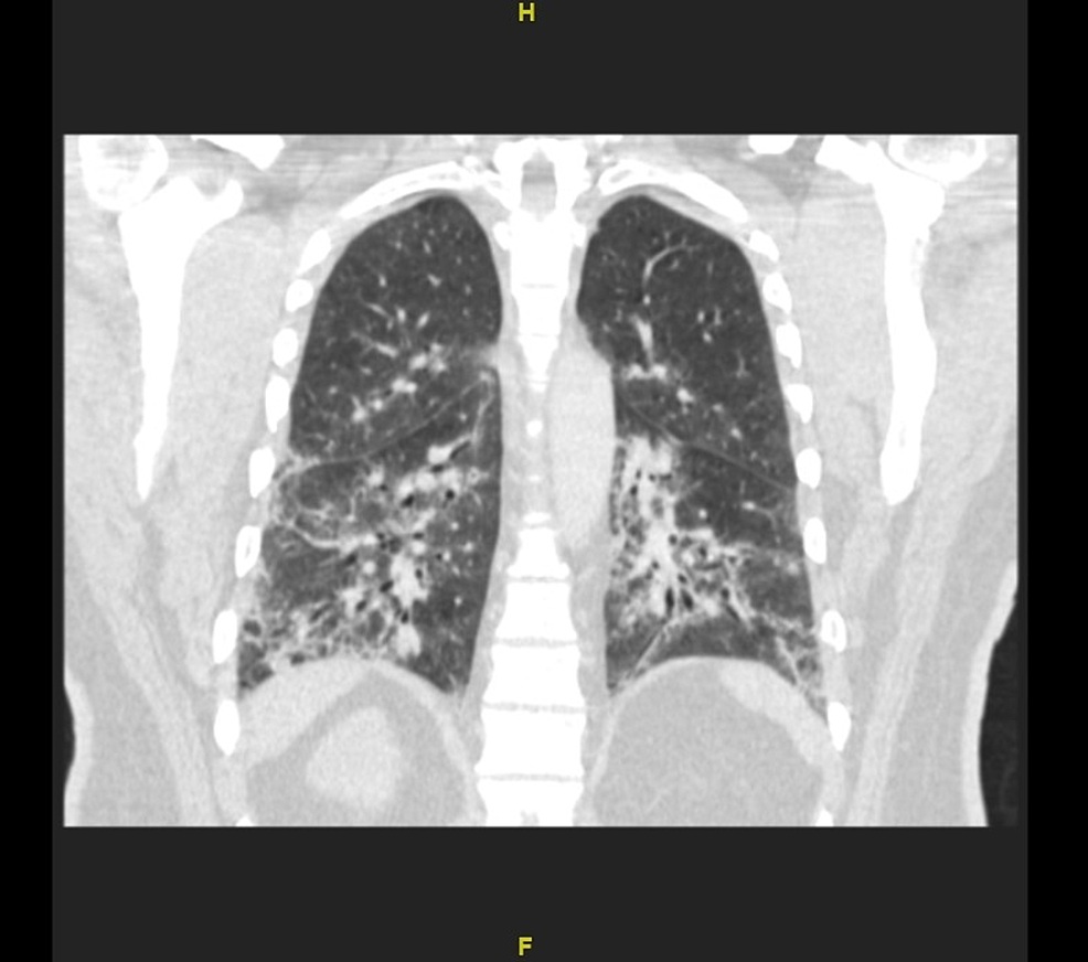
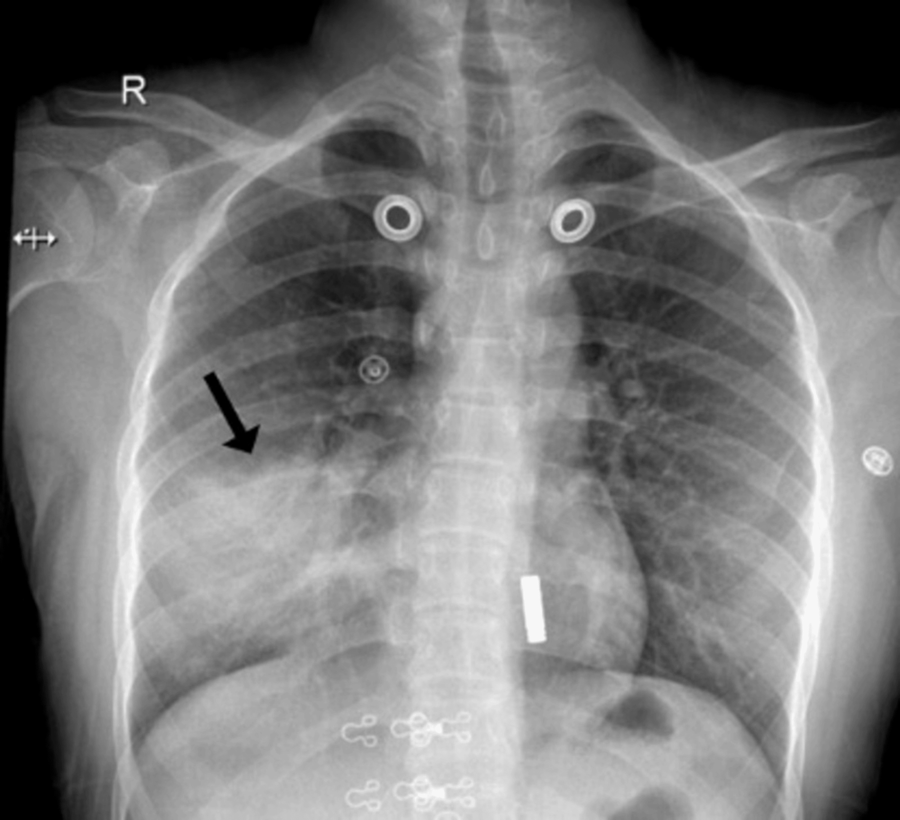
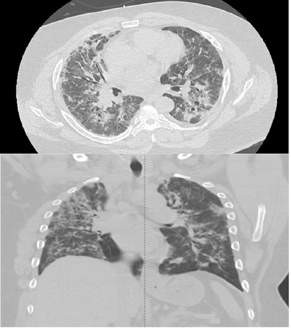
| HRCT — peripheral/subpleural consolidations | ~80% | Moderate (shared with eosinophilic pneumonia, vasculitis) | Bilateral, subpleural, and peribronchovascular areas of consolidation with or without GGO are the dominant CT finding in COP. The consolidations are often multifocal and can migrate from zone to zone on serial imaging — a feature that helps distinguish COP from malignancy and infection. Air bronchograms are commonly visible within consolidations. |
| Pulmonary function tests (PFTs) | High sensitivity for restriction | — | Mild to moderate restrictive defect (reduced TLC, FVC); DLCO is reduced. FEV₁/FVC ratio is normal or elevated. In a subset of patients (particularly those with small-airway involvement or pre-existing COPD), an obstructive or mixed pattern may be seen. Severity of PFT abnormality correlates roughly with radiographic extent of consolidation.[8] |
| BAL differential cell count | Moderate sensitivity (not required for diagnosis) | — | BAL in COP shows a characteristic mixed-cellularity pattern: elevated lymphocytes (often 20–40%), elevated neutrophils (~10–20%), and elevated eosinophils (~5%). This "dirty BAL" with elevations in all three cell lines contrasts with pure lymphocytosis (HP, NSIP) or pure neutrophilia (bacterial pneumonia) and helps support the diagnosis when combined with CT findings. BAL is most useful to exclude infection before starting corticosteroids. |
| Surgical lung biopsy (VATS/cryobiopsy) | Gold standard | High when combined with clinical/imaging context | Shows Masson bodies — intraluminal plugs of loose fibroblastic connective tissue in a myxoid matrix filling the alveolar lumen and respiratory bronchioles. Temporal uniformity (all lesions at the same stage) distinguishes COP from UIP (which shows temporal heterogeneity). Inflammatory infiltrate involves the surrounding alveolar walls. VATS biopsy is not always required when the CT pattern is typical and BAL excludes infection — clinical judgment applies. |
| Serologic workup (CTD panel) | Essential (not diagnostic of COP itself) | — | ANA with pattern, RF, anti-CCP, anti-Scl-70, myositis-specific antibodies (anti-Jo-1, anti-MDA5, anti-Mi-2), anti-SSA/SSB. COP presenting as the initial manifestation of polymyositis or dermatomyositis is well documented. Positive serology should prompt rheumatology consultation. Secondary COP from CTD requires treatment of both the OP and the underlying autoimmune disease. |
Cryptogenic organizing pneumonia produces a distinctive lung ultrasound signature that is both diagnostically informative and clinically underused. The dominant POCUS finding in COP is peripheral subpleural consolidation — hypoechoic, tissue-density regions abutting the pleural surface, often wedge-shaped or irregular in contour, with visible air bronchograms within the consolidation. These consolidations correspond directly to the patchy subpleural infiltrates seen on HRCT and are detectable by ultrasound with higher sensitivity than chest X-ray for peripheral lesions. In a patient presenting with cough, fever, and bilateral airspace disease that has not cleared on antibiotics, bedside lung ultrasound that reveals bilateral peripheral consolidations with dynamic air bronchograms — rather than the lobar, gravity-dependent consolidation of bacterial pneumonia — should shift the differential toward organizing pneumonia, eosinophilic pneumonia, or vasculitis.[10]
Dynamic vs. static air bronchogram: This is the single most important POCUS distinction in consolidation assessment. A dynamic air bronchogram — hyperechoic foci within a consolidation that move toward the probe with inspiration (centrifugal airflow) — is highly specific for pneumonia (bacterial or organizing). A static air bronchogram that does not move is more consistent with atelectasis. In COP, dynamic air bronchograms are typically present because the alveolar plugs of granulation tissue maintain airway patency and allow inspiratory airflow to reach the distal airways, producing the centrifugal bronchogram movement. Their presence in a patient with bilateral consolidations failing antibiotic therapy strengthens the case for organizing rather than resolving infectious pneumonia.
B-lines in COP: B-lines (vertical hyperechoic artifacts arising from the pleural line, reaching the far field without fading) may be present in zones adjacent to areas of consolidation in COP. They reflect partial alveolar flooding and interstitial thickening at the periphery of consolidating zones. Unlike the smooth, uniformly distributed bilateral B-lines of pulmonary edema, B-lines in COP are patchy, focal, and arise adjacent to areas of consolidation rather than from a smooth pleural line — tracking the same peripheral, non-gravitational distribution seen on HRCT.[11]
1. Bilateral peripheral subpleural consolidations with dynamic air bronchograms = organizing pneumonia until proven otherwise. When bilateral peripheral consolidations with dynamic air bronchograms persist after 2–3 antibiotic courses, lung ultrasound becomes the fastest bedside tool to confirm the pattern. The consolidations in COP are non-lobar, non-gravitational, and peripheral — not the posterior-dependent pattern of aspiration or the lobar pattern of Streptococcal pneumonia. Bringing the probe to areas of dullness on percussion or areas highlighted on CXR will reveal the consolidation pattern within minutes. A focused 8-zone scan of the lateral and posterior lung takes less than 5 minutes and can provide this information at the bedside before CT is ordered.
2. The reverse halo sign on ultrasound. While the reverse halo (atoll) sign is primarily a CT diagnosis, ultrasound-detected peripheral consolidations with surrounding hyperechoic B-line halos at the margins — corresponding to the rim of ground-glass opacity surrounding the central consolidation on CT — have been reported in COP. This pattern, while not yet formally validated in POCUS literature for COP diagnosis, is mechanistically plausible: the central consolidation appears as a hypoechoic tissue-density region while the surrounding ground-glass zone produces peripheral B-lines. Awareness of this potential ultrasound correlate is a clinical pearl for the experienced POCUS practitioner.
3. Serial POCUS for treatment response assessment. One of the most practical applications of POCUS in COP is monitoring the dramatic radiographic improvement that follows corticosteroid therapy. Subpleural consolidations that are visible and measurable at baseline should diminish substantially within 2–4 weeks of starting prednisone 40–60 mg/day. Serial POCUS at follow-up visits provides a radiation-free, real-time assessment of consolidation resolution — particularly useful in younger patients or those with high cumulative radiation exposure. Failure of consolidations to resolve or reduce in size at 4 weeks should prompt CT re-evaluation and consideration of alternative diagnoses or refractory disease.
4. Exclude pleural effusion and pulmonary embolism as competing diagnoses. COP can coexist with or be mimicked by conditions that also cause dyspnea and consolidation. POCUS pleural assessment (posterolateral thorax, patient seated) can rapidly identify pleural effusion, which is uncommon in COP but suggests infection, malignancy, or CTD-associated disease. POCUS for DVT (bilateral lower extremity compression) combined with right heart strain assessment (RV dilation, D-sign, McConnell's sign) should be performed when PE is in the differential — because the peripheral consolidation pattern of pulmonary infarction can closely mimic COP on both CXR and CT.
The laboratory workup in COP is primarily aimed at excluding competing diagnoses — infection, CTD, and malignancy — rather than establishing COP itself. Elevated inflammatory markers are nearly universal: ESR is typically markedly elevated (often >100 mm/h), CRP is elevated, and leukocytosis may be present. These findings often contribute to the initial misdiagnosis of bacterial pneumonia. CBC with differential, BMP, LFTs, and LDH provide baseline data and help assess disease severity. The landmark test is the HRCT chest.[1]
HRCT chest: The imaging cornerstone. Classic CT findings in COP include: (1) bilateral, patchy, subpleural and/or peribronchovascular consolidations and GGO — often with a lower-lung predominance but potentially affecting any zone; (2) the reverse halo (atoll) sign when present — a central GGO surrounded by a dense rim of consolidation — near-pathognomonic for OP; (3) air bronchograms within consolidations, often visible; (4) migratory pattern — consolidations appearing in new areas and clearing from prior areas on serial imaging, a finding unique to COP among the IIPs. Features that argue against COP: honeycombing, traction bronchiectasis (suggest UIP/NSIP), upper-lobe predominance of nodules (suggest HP or sarcoid), pleural-based masses (suggest malignancy or organizing effusion).[2]
Serologic evaluation: ANA (with reflex pattern), RF, anti-CCP, anti-Scl-70, myositis-specific antibodies (anti-Jo-1, anti-MDA5, anti-Mi-2), anti-SSA/SSB, anti-dsDNA, ANCA. Elevated CK and LDH raise concern for inflammatory myopathy (polymyositis/dermatomyositis-associated COP). A drug history is mandatory — any medication started in the months preceding symptom onset should be considered a potential causative agent.
Bronchoscopy and BAL: Recommended to exclude infection (bacterial, mycobacterial, fungal) before initiating corticosteroids. BAL differential shows mixed-cellularity pattern (lymphocytes, neutrophils, eosinophils all elevated). Transbronchial biopsy can show organizing pneumonia pattern but has limited sensitivity due to small sample size — a transbronchial biopsy showing OP supports the diagnosis but a negative biopsy does not exclude it. Bronchoalveolar lavage cytology may show lipid-laden macrophages in drug-induced OP.
Surgical lung biopsy: VATS or cryobiopsy is indicated when CT findings are atypical, when clinical progression is rapid, or when the differential includes malignancy (lymphoma, adenocarcinoma presenting as consolidation). Histologic demonstration of Masson bodies (intra-alveolar fibroblast plugs in a myxoid matrix) with temporal uniformity and preserved alveolar architecture establishes the definitive diagnosis. Perioperative risk in COP is substantially lower than in advanced IPF, given that COP patients typically have less underlying fibrosis and better baseline physiology.
| Intervention | When & How | Evidence & Key Points |
|---|---|---|
| Prednisone (initial high-dose) | Start at 0.75–1 mg/kg/day (typically 40–60 mg/day) for 4–8 weeks. Patients with moderate-to-severe disease or hypoxemia may begin at 1 mg/kg/day. Use the lowest effective dose for each individual patient — some mild cases respond to 0.5 mg/kg/day. Clinical and radiographic response is typically dramatic within 1–2 weeks. | Corticosteroids are the established first-line treatment for COP — no controlled trials exist for the initial response phase, but observational series consistently demonstrate response rates of 65–85% with prednisone.[8] The dramatic speed of improvement (consolidations visibly reducing within 2 weeks on HRCT) is itself diagnostically informative — slow or absent response should prompt reconsideration of the diagnosis. Monitor blood glucose, blood pressure, and bone protection from treatment initiation. |
| Prednisone taper (initial course) | After initial 4–8 weeks at therapeutic dose: reduce by 10 mg every 2–4 weeks to 20 mg/day; then reduce by 2.5–5 mg every 4 weeks thereafter. Total course: 6–12 months. Historically 6 months was standard; shorter 3-month courses have been trialed but carry higher relapse risk. Clinical and imaging reassessment before each dose reduction. | The relapse rate during taper is 13–58% across published series. Lazor et al. reported that 58% of 48 biopsy-proven COP patients relapsed, and 68% of first relapses occurred while still on treatment — meaning the drug was insufficient, not just withdrawn too early.[12] Predictors of relapse include disease in all three lung zones at baseline, high neutrophil percentage on BAL, and radiographic evidence of traction bronchiectasis. Slower tapers in high-risk patients (CTD-associated, bilateral disease, initial hypoxemia) reduce but do not eliminate relapse risk. |
| Relapse management | At relapse: re-escalate prednisone to the last effective dose (often the dose at which consolidations were controlled) and hold for 4 weeks, then taper more slowly than on the first course. For second relapse: use same re-escalation strategy; consider steroid-sparing agent. Most relapses respond promptly to dose re-escalation. Severe or hypoxemic relapse may require initial IV methylprednisolone 1–2 mg/kg/day for 3–5 days. | Relapses in COP do NOT carry the same prognosis as acute exacerbations of IPF. Long-term outcomes remain favorable even in multiply-relapsing patients — cumulative mortality from COP alone is low. The clinical challenge is steroid burden over years of relapsing-remitting disease. Total steroid exposure with repeated courses must be managed proactively with bone protection (calcium/vitamin D, bisphosphonate if high-dose course >3 months) and glycemic monitoring.[12] |
| Secondary cause elimination | Mandatory in all cases: stop any suspected causative drug, treat the underlying CTD, ensure infectious cause has been eradicated. For CTD-associated COP: optimize DMARDs (hydroxychloroquine, mycophenolate, azathioprine, rituximab) targeted at the underlying disease while tapering prednisone. | Secondary OP responds less reliably to corticosteroid taper alone unless the underlying cause is addressed. Drug-induced OP may resolve partially with drug discontinuation alone, though steroid acceleration is standard. CTD-associated COP may require longer prednisone courses and DMARD optimization to prevent relapse.[7] |
| Steroid-sparing agents (azathioprine, mycophenolate, cyclophosphamide, rituximab) | Indicated for: ≥2 relapses on standard prednisone taper; inability to taper below 10–15 mg/day without relapse; intolerance to corticosteroids; severe/refractory COP not responding to prednisone alone. Azathioprine 2–3 mg/kg/day or mycophenolate 1,000–1,500 mg BID are first-line steroid-sparing options. Rituximab reported in case series for refractory CTD-associated COP. | No RCT data for steroid-sparing agents in COP. Evidence is limited to case series and expert opinion. Macrolide antibiotics (azithromycin) have been reported to produce modest improvement in small series — a plausible mechanism given macrolides' anti-inflammatory properties beyond their antimicrobial spectrum — but macrolide monotherapy should not replace prednisone in moderate-to-severe disease.[6] |
| Observation without treatment (mild/asymptomatic disease) | In patients with minimal symptoms, normal SpO₂, and limited radiographic extent, watchful waiting with close follow-up (clinical assessment and HRCT at 4–6 weeks) is reasonable. Some cases of COP resolve spontaneously, particularly post-infectious forms. If symptoms progress or consolidations do not improve, initiate prednisone. | Spontaneous remission is documented in COP — particularly in mild post-infectious cases — though it is not predictable. In patients with significant dyspnea, hypoxemia, or bilateral extensive disease, delaying treatment risks progressive consolidation and potentially fibrosing transformation. The risk-benefit ratio of early treatment with low-to-moderate prednisone is favorable given the high response rate and generally good tolerability at standard doses. |
| Domain | ATS/ERS 2013 Position | Clinical Implication |
|---|---|---|
| Classification of COP | Acute/subacute IIP — distinct from chronic fibrosing IIPs (IPF, NSIP) and smoking-related IIPs. COP categorized alongside acute interstitial pneumonia (AIP) in the acute/subacute group. | COP's favorable prognosis and steroid responsiveness distinguish it from all other major IIPs. Do not treat COP like IPF — antifibrotics are not indicated, immunosuppression is the correct therapy. |
| Preferred terminology | COP (cryptogenic organizing pneumonia) is the preferred term. BOOP is discouraged to avoid confusion with bronchiolitis obliterans syndrome (BOS) in transplant recipients. | Use COP in documentation and communication. Clarify BOOP vs. BOS for patients and consultants — they are entirely different diseases. |
| Histopathologic definition | Intra-alveolar plugs of granulation tissue (Masson bodies) in alveolar ducts and surrounding alveoli; mild alveolar wall inflammation; temporal and spatial uniformity (all lesions at the same stage). | Temporal uniformity on biopsy distinguishes COP from UIP (temporal heterogeneity) — a critical distinction with major prognostic implications. Biopsies showing mixed COP + UIP pattern should be interpreted cautiously. |
| Multidisciplinary diagnosis | Diagnosis of COP should integrate clinical, radiographic, and (when available) histopathologic data — ideally through a multidisciplinary ILD conference. | HRCT showing classic COP pattern + BAL excluding infection may be sufficient for treatment without surgical biopsy in typical cases. Atypical cases (unifocal "mass-like" lesion, rapid progression, CT features of fibrosis) require tissue diagnosis. |
| COP vs. secondary OP | COP is reserved for organizing pneumonia without an identifiable trigger. Identical pathology in the setting of CTD, drugs, or infection is termed "secondary organizing pneumonia" — not COP. | A thorough search for secondary causes is mandatory before assigning the diagnosis of COP. The distinction matters for prognosis and for defining the correct treatment target. |
BOS = bronchiolitis obliterans syndrome; COP = cryptogenic organizing pneumonia; BOOP = bronchiolitis obliterans organizing pneumonia; IIP = idiopathic interstitial pneumonia; UIP = usual interstitial pneumonia; CTD = connective tissue disease.
The defining clinical problem in COP is relapse on steroid tapering. Lazor et al. demonstrated that 58% of 48 biopsy-proven COP patients relapsed during or after treatment.[12] More striking: 68% of first relapses occurred while patients were still receiving prednisone, often at doses between 10 and 20 mg/day — indicating that the relapse threshold for many patients is well above the doses at which most clinicians feel comfortable tapering. This finding has two practical implications: (1) taper should be driven by clinical and imaging response, not by a fixed timeline; and (2) the minimum effective maintenance dose (often 10–15 mg/day) is not negligible and must be weighed against the cumulative toxicity of prolonged low-dose corticosteroids in patients who may require years of treatment.
Despite the high relapse rate, the long-term prognosis of COP remains favorable — relapses respond to re-treatment, and COP-specific mortality is low. The key question for the fellow is whether a given patient's "COP" is truly idiopathic or whether there is an underlying CTD that has not yet declared itself clinically. Organizing pneumonia can precede the rheumatologic manifestations of dermatomyositis or anti-synthetase syndrome by months to years. Patients with COP should have their myositis-specific antibody panel checked (anti-Jo-1, anti-MDA5, anti-Mi-2, anti-PL-7, anti-PL-12) at baseline — and a negative panel does not rule out future CTD emergence. Follow these patients with a rheumatologist and re-screen serology annually in the first 3–5 years.
Focal organizing pneumonia (FOP) is a distinct entity from the multifocal COP described above. FOP presents as a solitary pulmonary nodule or mass — often found incidentally on imaging — and is typically asymptomatic. On CT, FOP is a peripheral, pleural-based consolidation that may have spiculated margins and air bronchograms, making it radiographically indistinguishable from early-stage lung cancer. Most FOP lesions are resected or biopsied for diagnostic purposes; the diagnosis is made histologically when the "mass" turns out to be a wedge-shaped plug of organizing pneumonia without features of malignancy. After surgical resection, recurrence is rare. FOP shares the same underlying pathology as diffuse COP but has a completely different clinical presentation, trajectory, and treatment strategy — reinforcing that the "COP" diagnosis should always specify whether it refers to the classic diffuse multifocal form or the focal solitary-lesion variant.
Epler GR et al. (n=50) — The landmark 1985 paper that coined the term BOOP and established the clinical phenotype: subacute flu-like illness, bilateral consolidations, steroid responsiveness, high relapse rate. The foundational reference for every COP clinician.
Cordier JF — The definitive comprehensive review of COP from the pioneer of European COP research. Covers pathology, CT features, clinical course, relapse biology, and treatment — the most-cited single reference on COP. Essential reading for any ILD fellow.
Lazor R et al. (n=48, biopsy-proven COP) — The primary evidence base for relapse management in COP. Demonstrated 58% relapse rate; 68% of first relapses occurred while still on treatment. Established the rationale for prolonged, slow steroid tapers in COP.
Drakopanagiotakis F et al. — Prospective comparison of cryptogenic vs. secondary OP in 168 patients. CTD-associated OP had longer treatment courses; both forms responded to steroids. Established clinical features distinguishing primary from secondary OP in a modern cohort.
[1] Hunninghake GM, Rosas IO. Interstitial Lung Disease. In: Jameson JL et al., eds. Harrison's Principles of Internal Medicine, 22nd ed. McGraw-Hill, 2025. (Harrison's Pulmonology, Ch. 304)
[2] Hager KM, Rubinowitz A, Gulati M. Idiopathic Interstitial Pneumonias Other Than Idiopathic Pulmonary Fibrosis. In: Grippi MA et al., eds. Fishman's Pulmonary Diseases and Disorders, 6th ed. McGraw-Hill, 2023. (Ch. 55)
[3] Davison AG, Heard BE, McAllister WAC, Turner-Warwick ME. Cryptogenic organizing pneumonitis. Q J Med. 1983;52(207):382–394. PMID: 6647749. [PubMed]
[4] Epler GR, Colby TV, McLoud TC, Carrington CB, Gaensler EA. Bronchiolitis obliterans organizing pneumonia. N Engl J Med. 1985;312(3):152–158. PMID: 3965933. [PubMed]
[5] Travis WD, Costabel U, Hansell DM, et al. An official ATS/ERS statement: update of the international multidisciplinary classification of the idiopathic interstitial pneumonias. Am J Respir Crit Care Med. 2013;188(6):733–748. PMID: 24032382. [PubMed]
[6] Cordier JF. Cryptogenic organising pneumonia. Eur Respir J. 2006;28(2):422–446. PMID: 16880372. [PubMed]
[7] Drakopanagiotakis F, Paschalaki K, Abu-Hijleh M, et al. Cryptogenic and secondary organizing pneumonia: clinical presentation, radiographic findings, treatment response, and prognosis. Chest. 2011;139(4):893–900. PMID: 20724743. [PubMed]
[8] Lohr RH, Boland BJ, Douglas WW, et al. Organizing pneumonia: features and prognosis of cryptogenic, secondary, and focal variants. Arch Intern Med. 1997;157(12):1323–1329. PMID: 9201006. [PubMed]
[9] Istrail L. The POCUS Textbook: Learn Point-of-Care Ultrasound of the Blood Vessels, Heart, & Lungs. ZeroGray Publishing, 2025. ISBN: 979-8-218-68730-4. [Buy on Amazon]
[10] Volpicelli G, Elbarbary M, Blaivas M, et al. International evidence-based recommendations for point-of-care lung ultrasound. Intensive Care Med. 2012;38(4):577–591. PMID: 22392031. [PubMed]
[11] Gargani L, Bruni C, Romei C, et al. Lung ultrasound B-lines in interstitial lung disease: moving from diagnosis to prognostic stratification. Chest. 2020;158(4):1515–1525. PMID: 32360727. [PubMed]
[12] Lazor R, Vandevenne A, Pelletier A, Leclerc P, Court-Fortune I, Cordier JF. Cryptogenic organizing pneumonia: characteristics of relapses in a series of 48 patients. Am J Respir Crit Care Med. 2000;162(2):571–577. PMID: 10934089. [PubMed]